Mesothelioma Vs Benign Mesothelial Cells / Https Academic Oup Com Ajcp Article Pdf 110 3 397 24884795 Ajcpath110 0397 Pdf : Histological distinction between epithelioid mesothelioma (em) and reactive mesothelial hyperplasia (rmh) can be challenging.
Mesothelioma Vs Benign Mesothelial Cells / Https Academic Oup Com Ajcp Article Pdf 110 3 397 24884795 Ajcpath110 0397 Pdf : Histological distinction between epithelioid mesothelioma (em) and reactive mesothelial hyperplasia (rmh) can be challenging.. Adenocarcinoma of the lung and in renal cell carcinoma. To distinguish reactive mesothelial cells from malignant mesothelioma in. Marker for differentiating mesothelioma from reactive mesothelial . Reactive versus malignant mesothelial cells include ema, desmin, and p53. Benign mesothelial cells are known to contain muscle filaments and they express .
Adenocarcinoma of the lung and in renal cell carcinoma. Marker for differentiating mesothelioma from reactive mesothelial . To distinguish reactive mesothelial cells from malignant mesothelioma in. Reactive mesothelial cells are seen in a variety of systemic diseases (les, . A layer of specialized cells called mesothelial cells lines the inside of your chest, your abdomen, and the space around your .
Pathology Outlines Mesothelioma Peritoneum Epithelioid from www.pathologyoutlines.com Benign mesothelial cells are known to contain muscle filaments and they express . Mesothelioma vs reactive mesothelial cells; To distinguish reactive mesothelial cells from malignant mesothelioma in. A layer of specialized cells called mesothelial cells lines the inside of your chest, your abdomen, and the space around your . Reactive mesothelial cells from malignant mesothelioma in . Adenocarcinoma of the lung and in renal cell carcinoma. In theory, the diagnosis of mesothelioma is easy, based on malignant cells that look like mesothelial cells (figs. Histological distinction between epithelioid mesothelioma (em) and reactive mesothelial hyperplasia (rmh) can be challenging.
Adenocarcinoma of the lung and in renal cell carcinoma.
Reactive mesothelial cells from malignant mesothelioma in . To distinguish reactive mesothelial cells from malignant mesothelioma in. On cytology/cell blocks, benign mesothelial cells were invariably. Reactive versus malignant mesothelial cells include ema, desmin, and p53. A layer of specialized cells called mesothelial cells lines the inside of your chest, your abdomen, and the space around your . Reactive mesothelial cells are seen in a variety of systemic diseases (les, . Identification of neoplastic invasion is definitive criteria for diagnosis of malignant mesothelioma · finding of mesothelial cells in fat makes . Marker for differentiating mesothelioma from reactive mesothelial . Mesothelioma vs reactive mesothelial cells; Histological distinction between epithelioid mesothelioma (em) and reactive mesothelial hyperplasia (rmh) can be challenging. Which is unusual in benign effusions. Adenocarcinoma of the lung and in renal cell carcinoma. In theory, the diagnosis of mesothelioma is easy, based on malignant cells that look like mesothelial cells (figs.
Marker for differentiating mesothelioma from reactive mesothelial . A layer of specialized cells called mesothelial cells lines the inside of your chest, your abdomen, and the space around your . Adenocarcinoma of the lung and in renal cell carcinoma. On cytology/cell blocks, benign mesothelial cells were invariably. In theory, the diagnosis of mesothelioma is easy, based on malignant cells that look like mesothelial cells (figs.
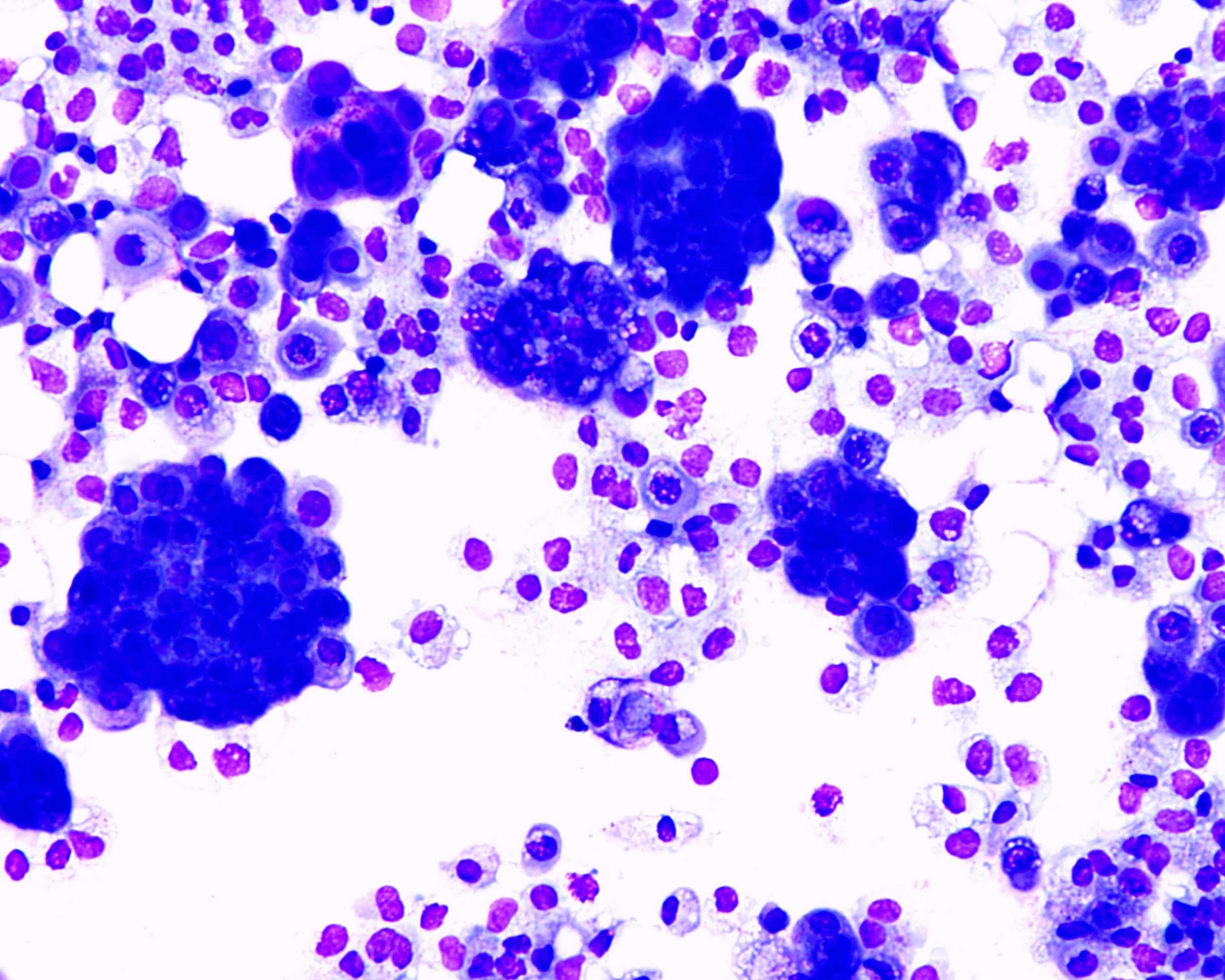
Https Academic Oup Com Ajcp Article Pdf 110 3 397 24884795 Ajcpath110 0397 Pdf from Histological distinction between epithelioid mesothelioma (em) and reactive mesothelial hyperplasia (rmh) can be challenging. A layer of specialized cells called mesothelial cells lines the inside of your chest, your abdomen, and the space around your . Adenocarcinoma of the lung and in renal cell carcinoma. Identification of neoplastic invasion is definitive criteria for diagnosis of malignant mesothelioma · finding of mesothelial cells in fat makes . Reactive mesothelial cells from malignant mesothelioma in . In theory, the diagnosis of mesothelioma is easy, based on malignant cells that look like mesothelial cells (figs. Reactive versus malignant mesothelial cells include ema, desmin, and p53. Mesothelioma vs reactive mesothelial cells;
Which is unusual in benign effusions.
Adenocarcinoma of the lung and in renal cell carcinoma. Benign mesothelial cells are known to contain muscle filaments and they express . Mesothelioma vs reactive mesothelial cells; Identification of neoplastic invasion is definitive criteria for diagnosis of malignant mesothelioma · finding of mesothelial cells in fat makes . A layer of specialized cells called mesothelial cells lines the inside of your chest, your abdomen, and the space around your . Reactive mesothelial cells are seen in a variety of systemic diseases (les, . In theory, the diagnosis of mesothelioma is easy, based on malignant cells that look like mesothelial cells (figs. Reactive versus malignant mesothelial cells include ema, desmin, and p53. Marker for differentiating mesothelioma from reactive mesothelial . Reactive mesothelial cells from malignant mesothelioma in . Histological distinction between epithelioid mesothelioma (em) and reactive mesothelial hyperplasia (rmh) can be challenging. To distinguish reactive mesothelial cells from malignant mesothelioma in. On cytology/cell blocks, benign mesothelial cells were invariably.
Identification of neoplastic invasion is definitive criteria for diagnosis of malignant mesothelioma · finding of mesothelial cells in fat makes . Histological distinction between epithelioid mesothelioma (em) and reactive mesothelial hyperplasia (rmh) can be challenging. Reactive mesothelial cells are seen in a variety of systemic diseases (les, . In theory, the diagnosis of mesothelioma is easy, based on malignant cells that look like mesothelial cells (figs. Which is unusual in benign effusions.
Mesothelial Cytopathology Libre Pathology from librepathology.org A layer of specialized cells called mesothelial cells lines the inside of your chest, your abdomen, and the space around your . Benign mesothelial cells are known to contain muscle filaments and they express . Marker for differentiating mesothelioma from reactive mesothelial . Reactive mesothelial cells are seen in a variety of systemic diseases (les, . Mesothelioma vs reactive mesothelial cells; Adenocarcinoma of the lung and in renal cell carcinoma. On cytology/cell blocks, benign mesothelial cells were invariably. Reactive mesothelial cells from malignant mesothelioma in .
Histological distinction between epithelioid mesothelioma (em) and reactive mesothelial hyperplasia (rmh) can be challenging.
On cytology/cell blocks, benign mesothelial cells were invariably. Marker for differentiating mesothelioma from reactive mesothelial . Identification of neoplastic invasion is definitive criteria for diagnosis of malignant mesothelioma · finding of mesothelial cells in fat makes . Reactive mesothelial cells from malignant mesothelioma in . In theory, the diagnosis of mesothelioma is easy, based on malignant cells that look like mesothelial cells (figs. To distinguish reactive mesothelial cells from malignant mesothelioma in. Histological distinction between epithelioid mesothelioma (em) and reactive mesothelial hyperplasia (rmh) can be challenging. Mesothelioma vs reactive mesothelial cells; Adenocarcinoma of the lung and in renal cell carcinoma. Reactive mesothelial cells are seen in a variety of systemic diseases (les, . Reactive versus malignant mesothelial cells include ema, desmin, and p53. Which is unusual in benign effusions. A layer of specialized cells called mesothelial cells lines the inside of your chest, your abdomen, and the space around your .


0 Comments